Medical imaging
Kidney stone segmentation in CT
Can a model delineate small kidney stones more accurately by narrowing the search before segmentation?
Biomedical Signal Processing and Control · 2026
Approach
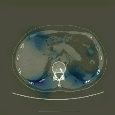
Two classifiers first identify kidney-containing slices and then stone-containing slices. The kidney stone segmentation network, KSSNet, processes the selected images and reuses features learned from kidney tissue segmentation.
Reported results
Table 7 shows the effect of adding the two-stage slice selection to the same KSSNet segmentation model.
- Dice with full scope reduction
- 97.44%
- Dice without scope reduction
- 90.13%
- Subjects in the source CT cohort
- 259
My contribution
I designed the successive scope-reduction and feature-transfer approach and led model development, experiments, analysis, and manuscript preparation.
Study scope
The five folds separate subjects. The scans came from a single hospital, so the reported scores describe that cohort and protocol.